Subjects
Grades
During cellular respiration, the cells of the body use oxygen |(\text{O}_2)| and glucose |(\text{C}_6\text{H}_{12}\text{O}_6),| and release carbon dioxide |(\text{CO}_2)| and water |(\text{H}_2\text{O})|. The cells perform a slow combustion reaction.
The role of the respiratory system is to supply the body with oxygen |(\text{O}_2)| and to expel carbon dioxide |(\text{CO}_2).|
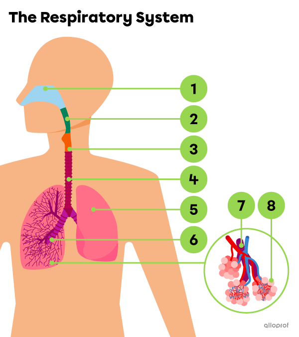
The main structures of the respiratory system are identified in the image below.
There are other structures closely related to the respiratory system. The pleura, diaphragm, intercostal muscles, ribs and sternum are involved in the respiratory movements that enable inhaling and exhaling.
Moments in the video:
| Description | Functions |
|---|---|
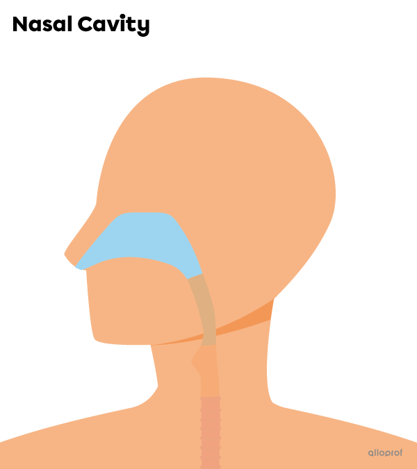
| The nasal cavity has two highly vascularized sections, lined with a thick mucous membrane, ciliated cells and hair. |
|
Although it’s possible to breathe through the mouth, the nasal cavity is still the most suitable structure for drawing air into the body.
Ciliated cells should not be confused with nose hair. The hair is clearly visible to the naked eye at the entrance to the nostrils. Ciliated cells are microscopic.

Nose hair at the entrance of the nostrils
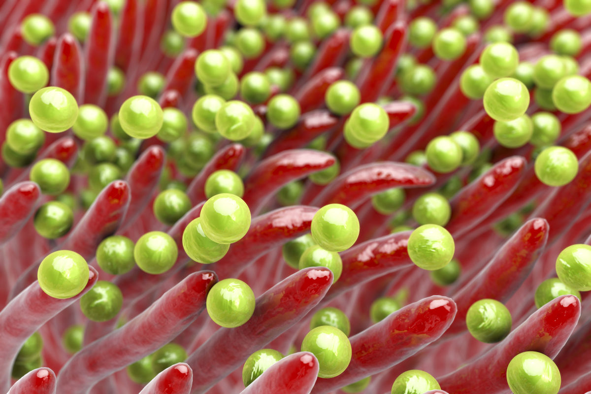
Bacteria (green spheres) trapped by the ciliated cells of the nasal cavity (red projections)
The air inhaled by the body contains fine dust, pollution particles and some allergens such as pollen. Several structures in the respiratory system are lined with a mucous membrane composed of ciliated cells and mucus-producing cells.
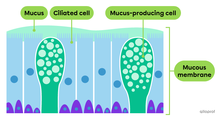
Illustration of the ciliated mucous membrane of the nasal cavity
The mucous membranes produce mucus, a viscous liquid that traps small, unwanted particles from the outside air. These membranes line the respiratory tract and become thinner between the nasal cavity and the alveoli. In the nasal cavity, the thick mucous membrane is responsible for humidifying the inhaled air.
The pharynx and the upper part of the larynx are lined with a very thin mucous membrane because these structures are exposed to abrasion by ingested food.
Ciliated cells, or cilia, help move mucus through the respiratory tract and trap particles. The ciliated cells of the trachea can propel mucus upward to be swallowed or coughed up. There are fewer ciliated cells between the nasal cavity and the bronchioles and none in the alveoli.
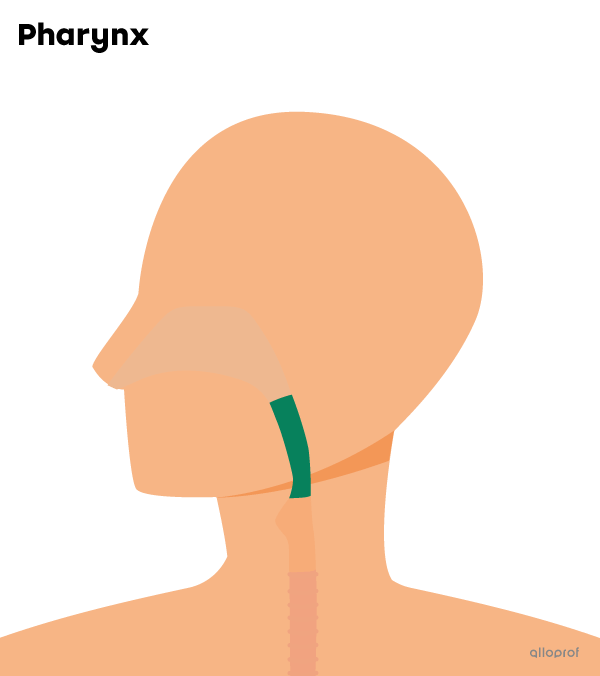
| Description | Function |
|---|---|
| The pharynx is the common tract between the respiratory system and the digestive system. | It ensures the passage of air between the nasal cavity and the larynx. |
The pharynx contains a muscle called the uvula, which prevents food from going up into the nasal cavity.
The pharynx also contains the tonsils, a structure of the lymphatic system. They produce antibodies to prevent and cure infections.
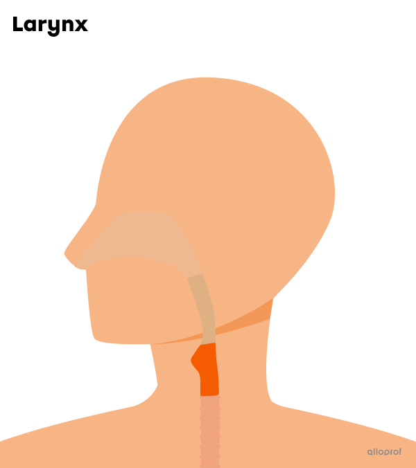
| Description | Function |
|---|---|
| The larynx is a cylindrical, cartilage tract. | It ensures the passage of air between the pharynx and the trachea. |
The larynx contains a cartilage structure called the epiglottis, which prevents food from entering the trachea.
The larynx also contains the vocal cords, which produce vocal sounds.
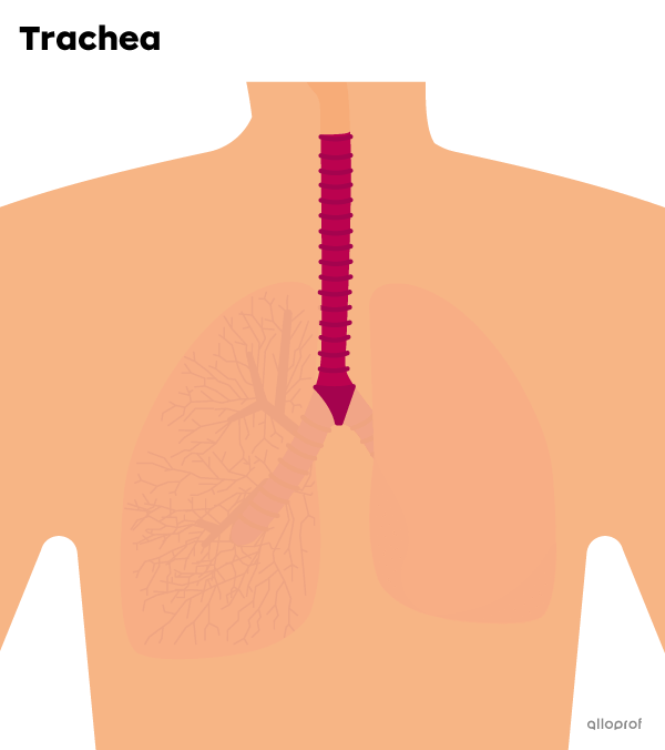
| Description | Function |
|---|---|
| The trachea is a flexible tube with rigid cartilage rings and ciliated cells. | It ensures the passage of air between the larynx and the bronchi. |
The cartilage rings keep the trachea open and prevent it from collapsing, which would prevent air flow.
The ciliated cells in the trachea move the mucus and any unwanted particles upward. This way, the mucus can be coughed up or swallowed.
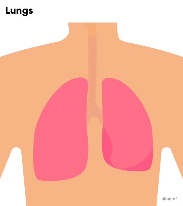
| Description | Functions |
|---|---|
| The lungs are two spongy, elastic organs. |
|
The left lung is smaller than the right lung to allow more room for the heart, which is positioned slightly to the left.
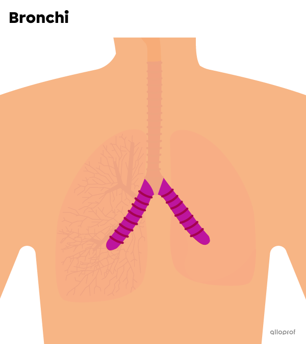
| Description | Function |
|---|---|
| The bronchi (singular: bronchus) are two ducts with rigid rings of cartilage leading to each lung. | They ensure the passage of air between the trachea and the bronchioles. |
The rings of cartilage keep the bronchi open and prevent them from collapsing, which would prevent air flow.
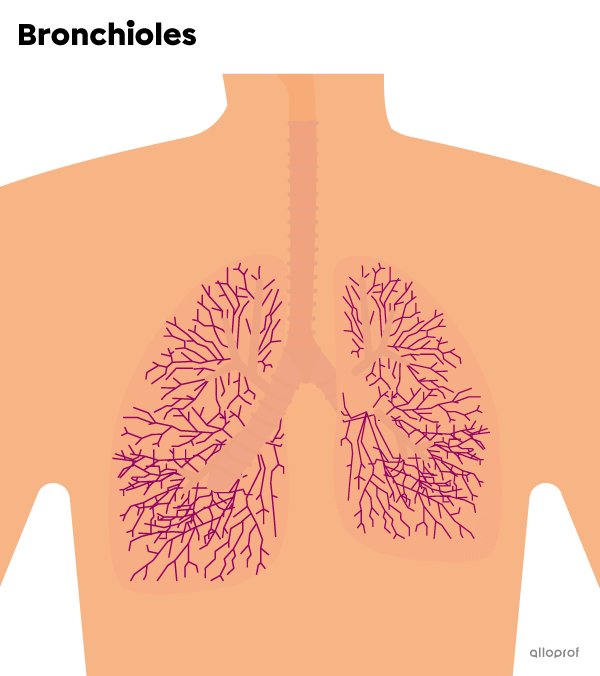
| Description | Function |
|---|---|
| The bronchioles are smaller ducts that branch off from the bronchi. | They ensure the air passage between the bronchioles and the alveoli. |
| Description | Fonction |
|---|---|
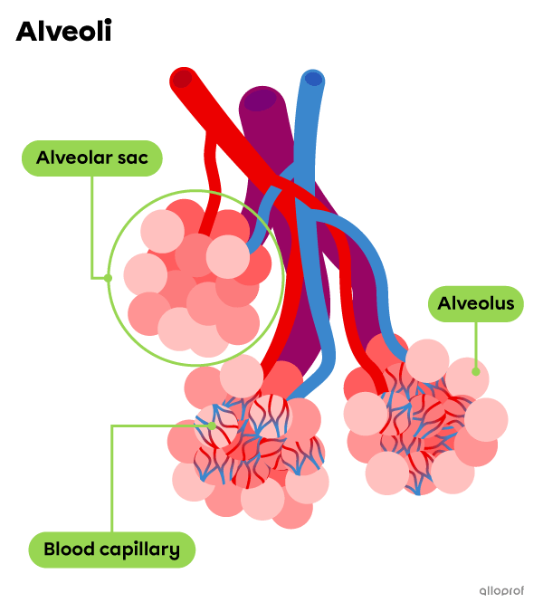
| The alveoli (singular: alveolus) are small bubbles at the ends of the bronchioles with thin walls that are permeable to gas. | They ensure gas exchanges between the air and bloodstream. |
The alveoli are grouped into alveolar sacs, covered with blood capillaries.